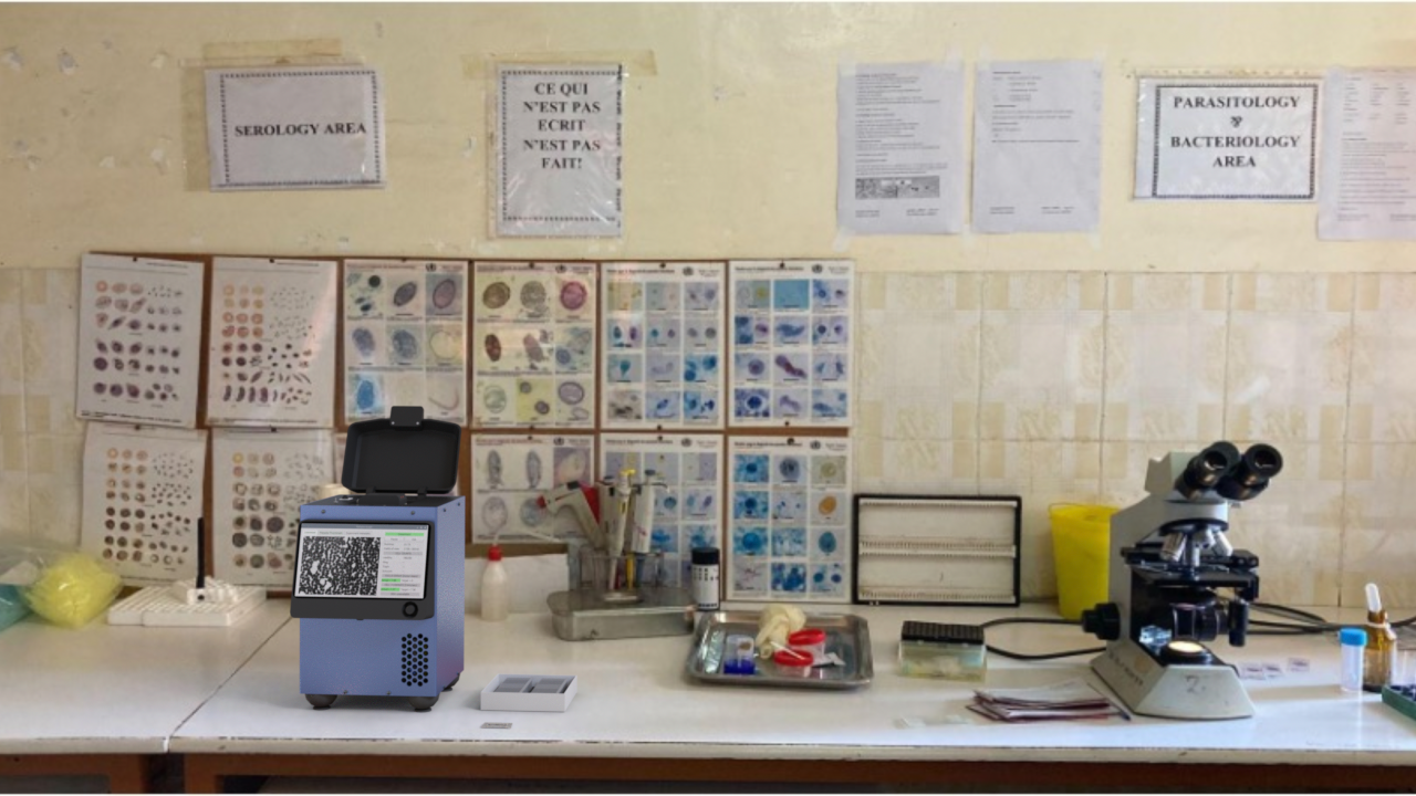
Across research, pharmaceutical, diagnostics, and clinical settings, the same bottleneck holds: sample prep, staining, manual microscopy, and specialist expertise.
Remoscope is one platform: a high throughput label-free imaging cytometer that classifies, counts, and learns from every cell it sees. It images millions of cells per sample without labels or sample preparation and analyzes morphology in real time. Models run on device, built for the lab, clinic, or field.
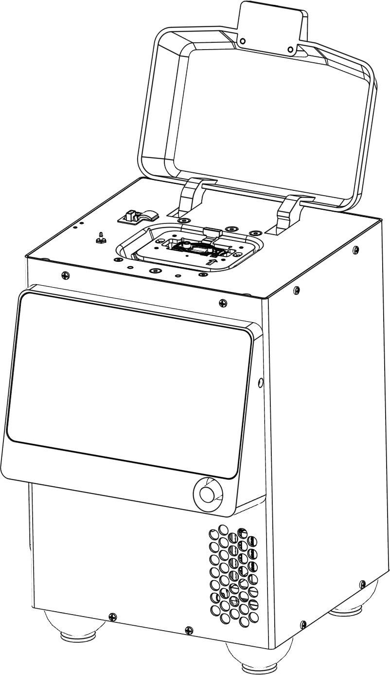
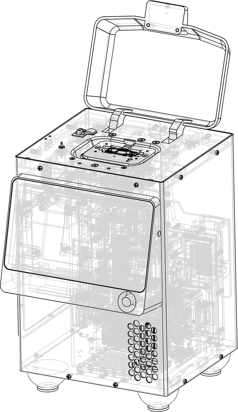
5–100× faster than conventional workflows
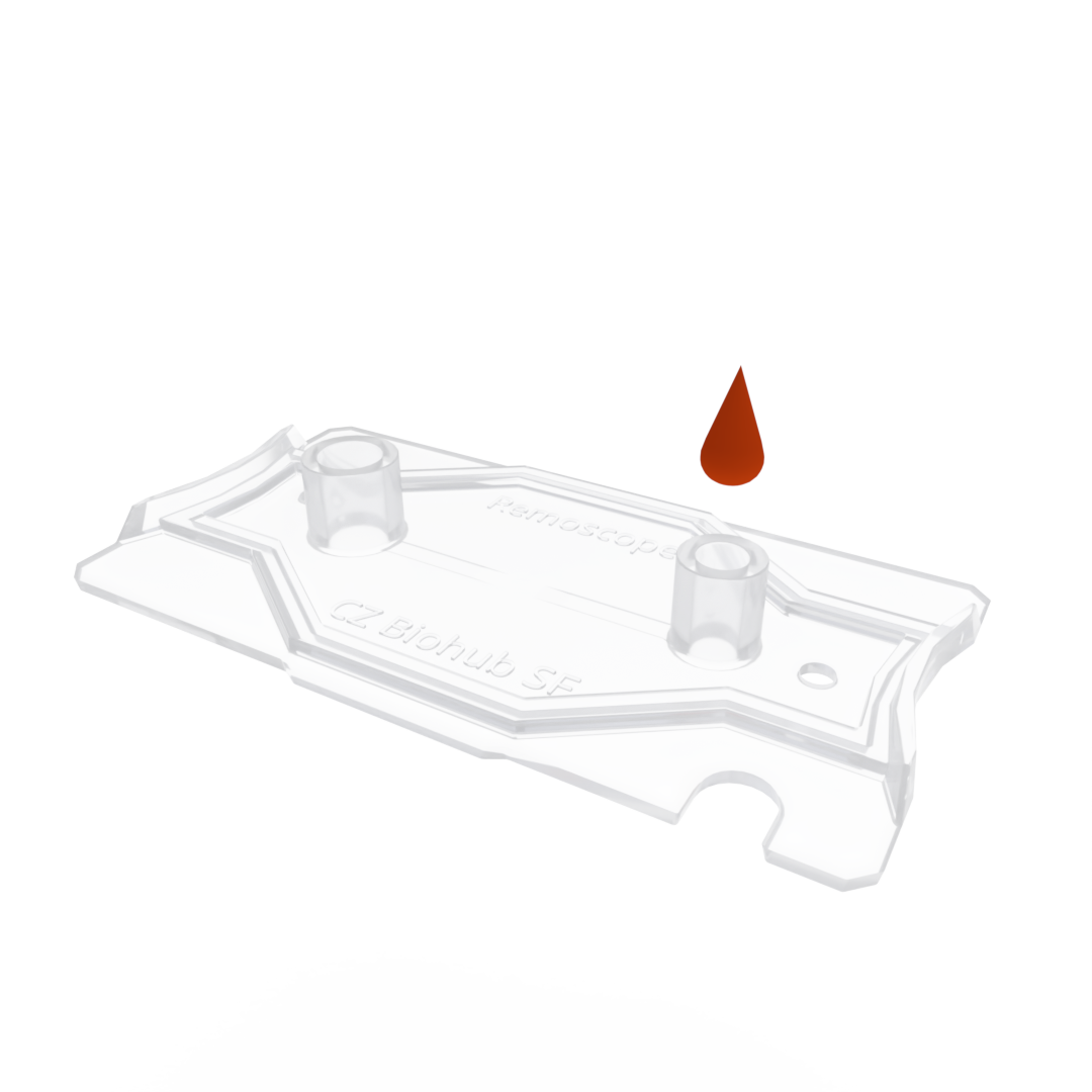
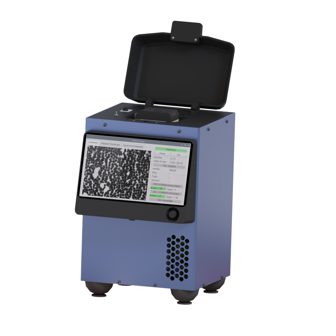

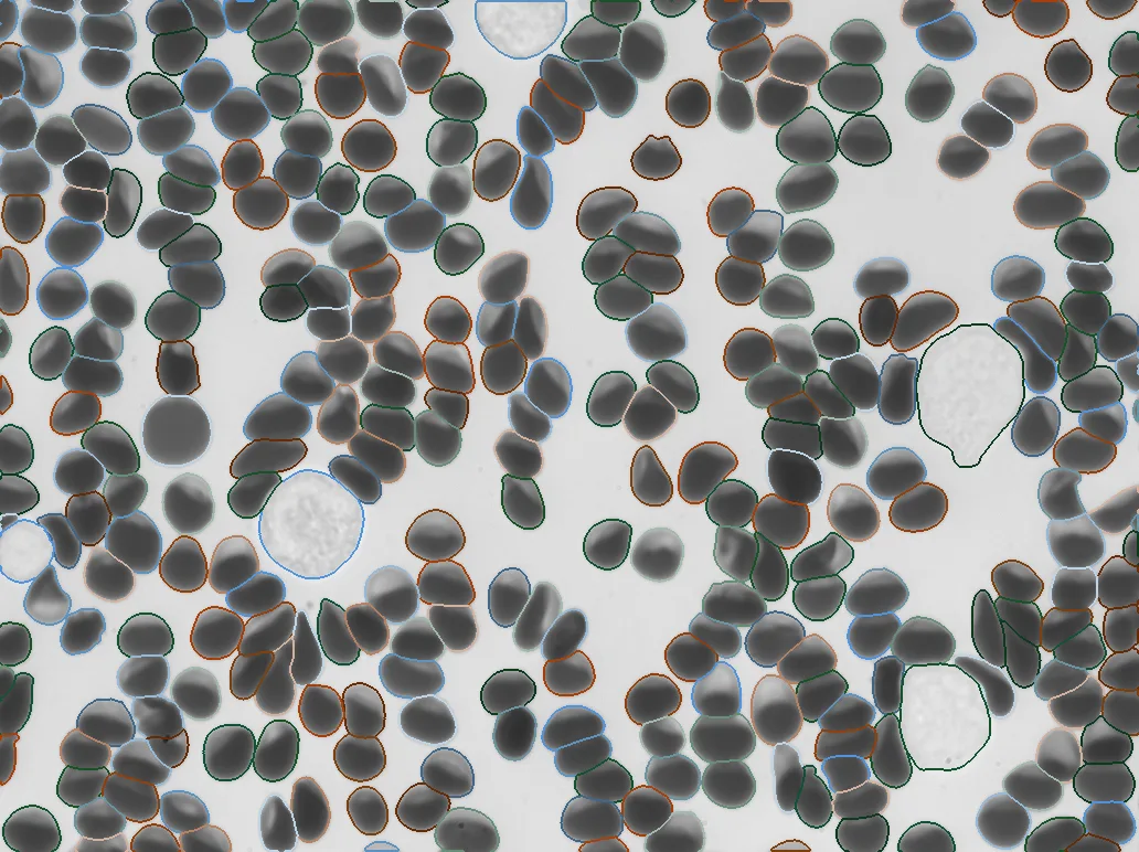
Every Remoscope generates standardized, labeled imaging data at scale. We build our own AI/ML foundation models which are fine-tuned for specific tasks like cell counts, subtype classification, anomaly detection, and diagnostics.
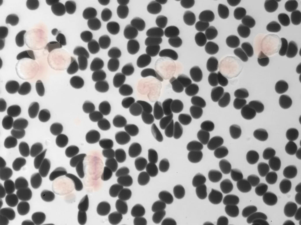
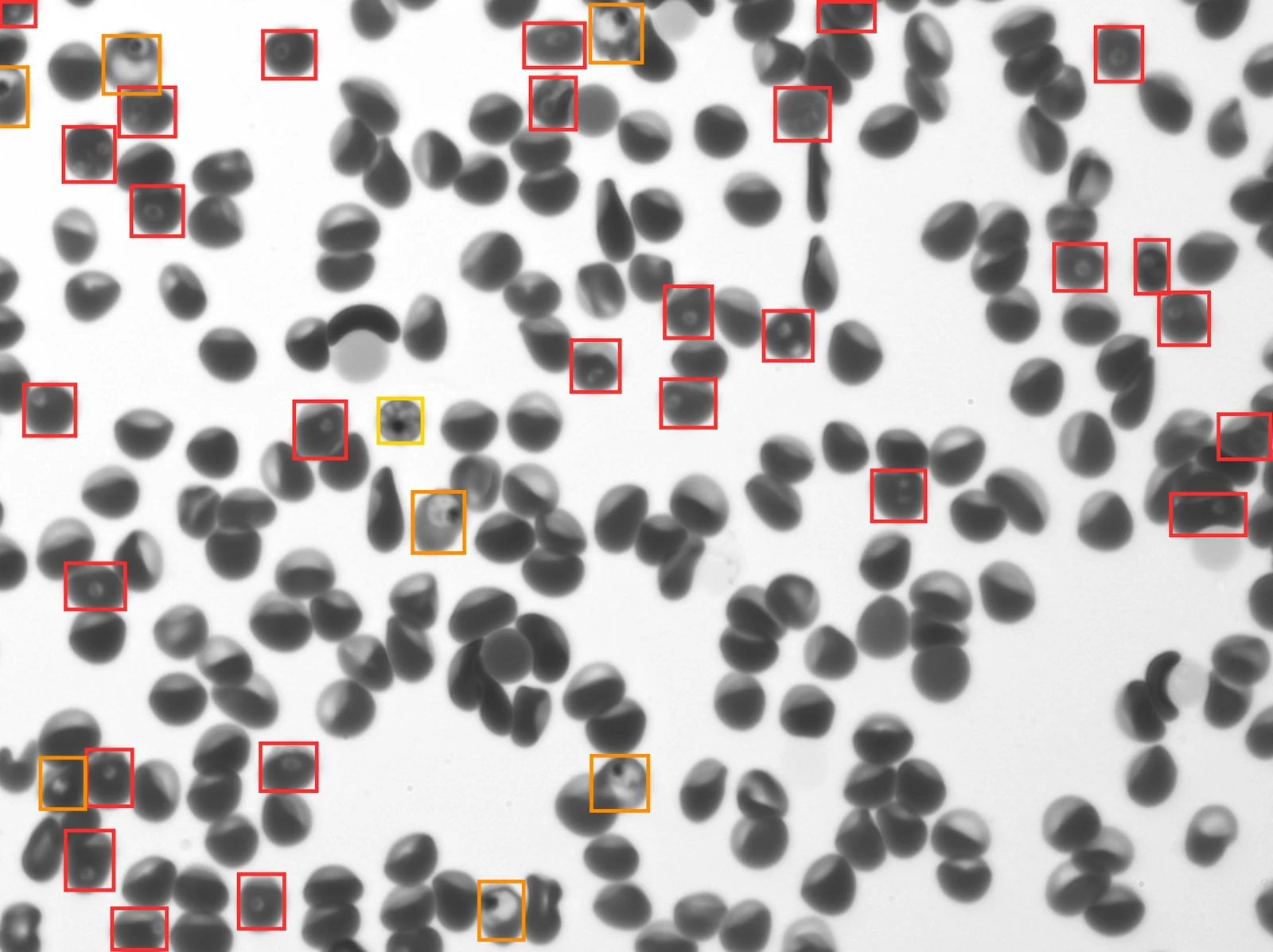
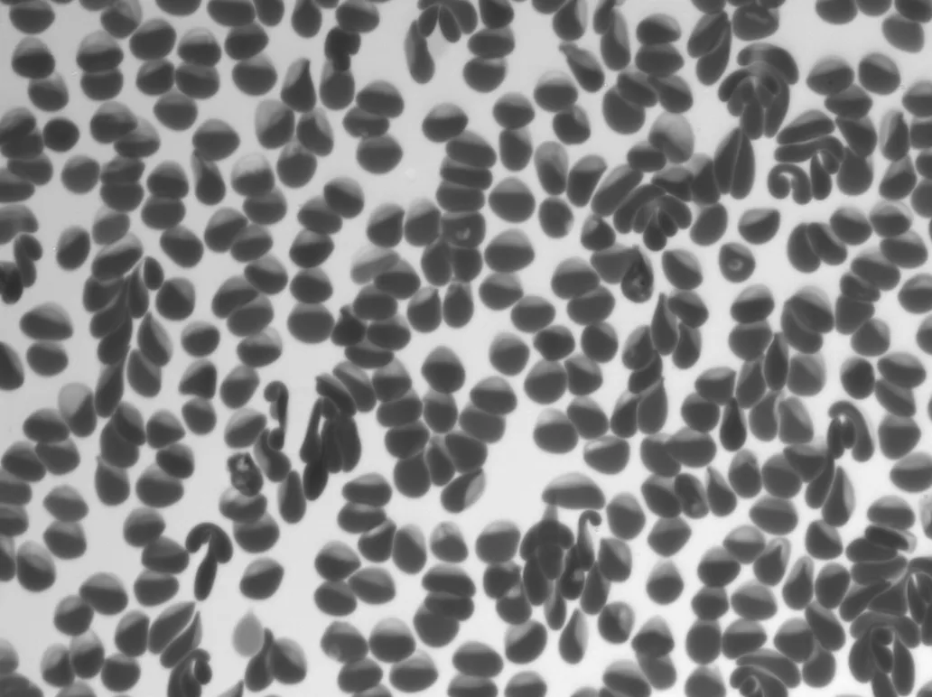
Quantifies Plasmodium falciparum parasites by life stage in fresh, undiluted whole blood in just 12 minutes, with no staining or fixation.
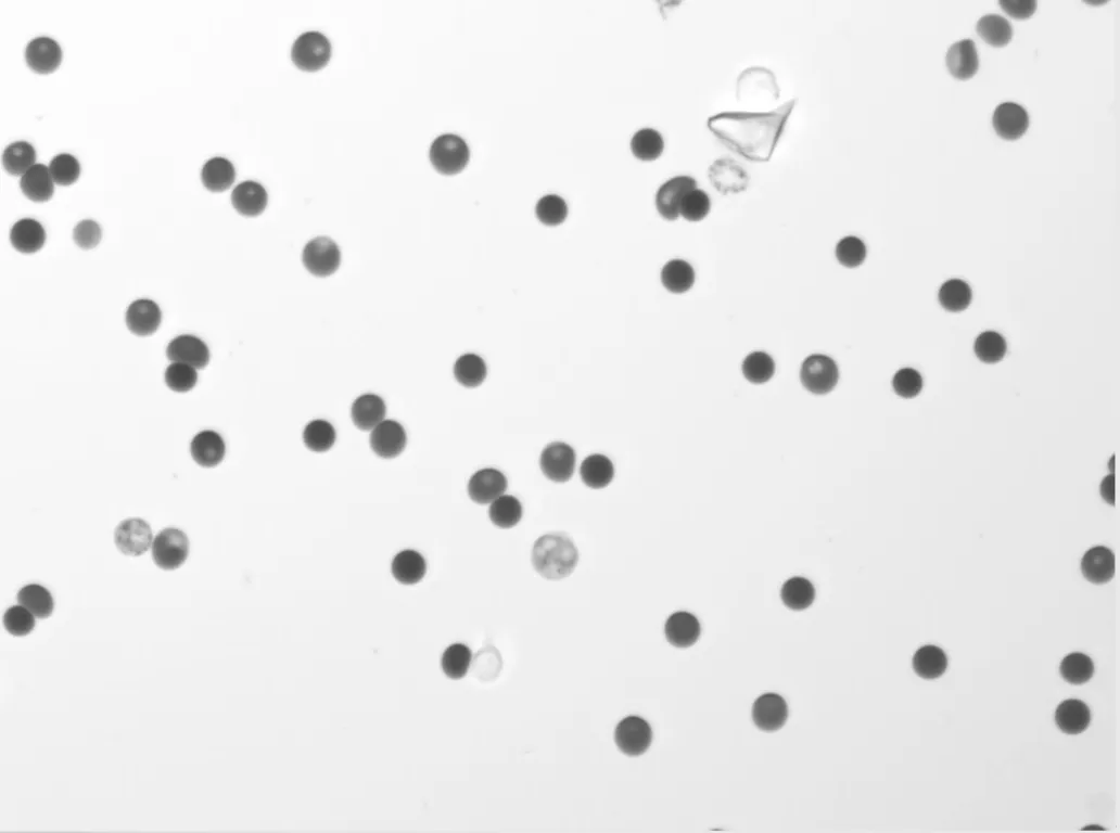
Due to its small sample volume, Remoscope is suited to veterinary and preclinical work, where small animal blood volumes limit conventional analyzers.
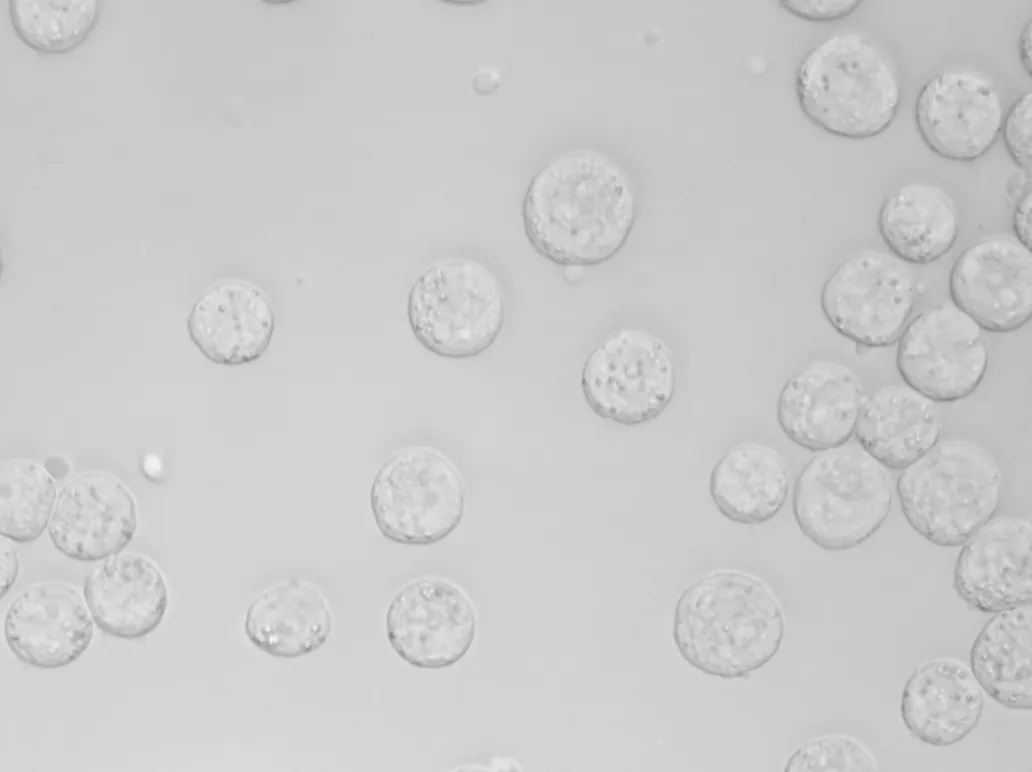
Distinguishes healthy, stressed, and dying cells in culture by morphology alone. Its small sample volume lets it integrate seamlessly into established cell-culture workflows.
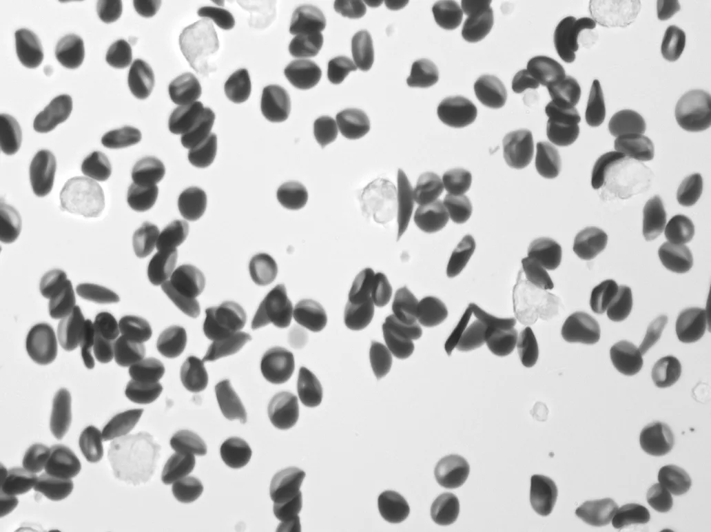
Detects sickled red blood cell morphology for screening and monitoring.
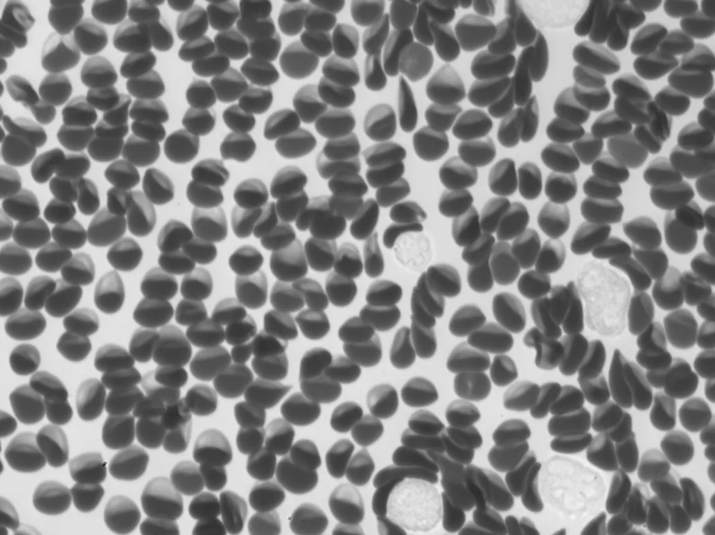
Distinguishes sepsis patients from other critically ill patients and healthy donors, from fresh undiluted whole blood in 12 minutes.
Supports rapid analysis of low-cellularity CSF samples that are slow to process by hand and otherwise require high-cost lab equipment. Screens for neurological immune conditions such as encephalitis and meningitis.
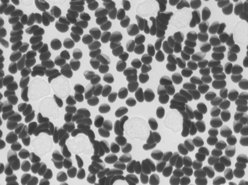
Surfaces abnormal white cell populations, like those seen in chronic myelomonocytic leukemia (CMML), from whole blood by morphology-based classification.
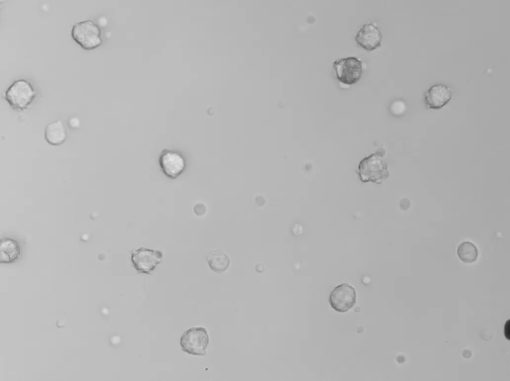
Uses label-free imaging to detect activated immune cells, enabling functional assays without antibody panels or flow-cytometry staining.
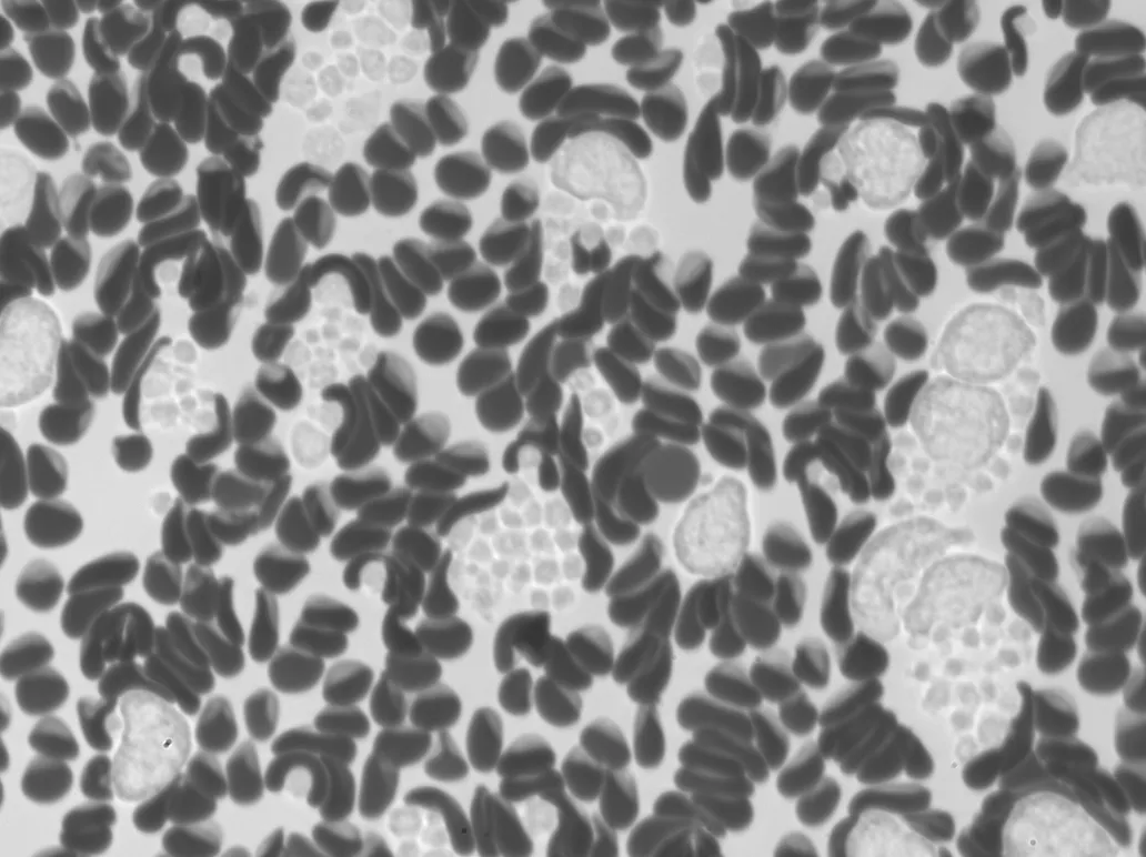
Able to flag abnormalities in whole blood, such as thrombocytosis, extending Remoscope to broad hematologic screening.
Every sample is classified label-free, in real time — on the same hardware.
The Remoscope is robust and repeatable, enabling both discovery and research-use-only clinical applications.
Feel free to book a demo, initiate a collaboration, or fill out a pre-order form!